Photoemission Electron Microscope (PEEM) for time-resolved ARPES experiments

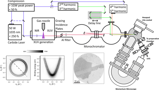
Our PEEM lab is equipped with a commercial photoemission electron microscope from SPECS GmbH (commercial name of the instrument: KREIOS). In the PEEM instrument, an immersion lens column generates an image of the lateral distribution of the photoelectrons I (x,y) and an additional Fourier lens transforms it into an image of the photoelectron emission angles. By an additional lens, either one of the two planes can be projected onto the detector. The latter contains the momentum distribution of the photoelectrons I (kx,ky) with high angular resolution. The photoelectrons are filtered by an hemispherical energy analyzer, which allows to acquire energy- and momentum-resolved two-dimensional (2D) maps at a fixed, and selectable, kinetic energy I (E,kx,ky). In this way it is possible to directly measure the spectral function of selected materials, which for non-correlated materials corresponds to the surface projected band structure.
Furthermore, our microscope offers up to five different field-of-views varying in diameter from 100 µm to 10 µm and from 6 Å-1 to 0.5 Å-1, in real and momentum operation modes, respectively. A piezo sample stage with 6 degrees of freedoms (for tilt and positioning) is coupled with an additional stage that allows to azimuthally rotate the sample ±90°, whereas a flow cryostat keeps the sample at 8 K, when cooled with liquid Helium. The extraordinary performance of our PEEM are demonstrate by measuring the photoelectron momentum distribution map for the Au(111) surface at the Fermi energy. The Shockley surface state is evident, and manifests as two concentric rings around Γ. On Au(111) the splitting of the surface state is a well-known phenomenon: the breaking of the inversion symmetry at the interface together with strong atomic spin-orbit coupling leads to the separation of the surface state in two branches with opposite spin-character (Rashba splitting). By fitting a line profile of the surface states with a Voigt function, we obtain a k resolution of 0.005 Å-1, which is comparable with state-of-the-art PEEMs with a double hemispherical analyzer. As a built-in excitation source, the microscope is equipped with a UVS 300 duoplasmatron type discharge VUV light source, yielding 21.2 eV photon energy, when He is employed.
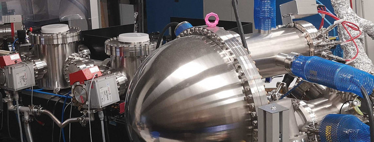
A home-assembled high harmonic generation (HHG) source is also coupled with the instrument. The seeding laser is a commercial Carbide (50 W, 1030 nm) from Light Conversion while the generation chamber is provided by Active Fiber Systems GmbH. To generate the HHG radiation we use an Ar gas stream jet with a nozzle of 150 µm diameter. A monochromator (Indigo Optical Systems GmbH) based on a multilayer mirror scheme filters the 25th harmonic providing a linearly-horizontal polarized light with an energy of 29.8 eV and a pulse duration of 300 fs with a repetition rate of 200 kHz.
The KREIOS is connected to a preparation chamber equipped with state-of-the-art surface characterization techniques, such as a Low Energy Electron Diffraction (LEED) instrument, a sputter gun and a self-standing AUGER electron spectrometer. A cluster flange allows to evaporate metals, by e-beam heating, while acquiring Medium Energy Electron Diffraction (MEED) patterns. A gate-valve coupled with a z-translator allows a fast exchange of the evaporants, as well as of the evaporator source without breaking the Ultra High Vacuum conditions (UHV better than 1∙10-10 mbar). A commercial KENTAX evaporator, for evaporating molecular compounds that may disrupt the cleanness of the preparation chamber, is mounted on an adjacent chamber (UHV better than 5∙10-10 mbar) separated by a gate valve.


